Mitral Annular Disjunction (MAD) And Refractory Ventricular Fibrillation
A 45-year-old male was followed in an outpatient clinic with a moderate mitral valve regurgitation (prolapse of both cusps) and mitral annular disjunction (MAD).
Transthoracic echocardiography documented:
• Normal ejection fraction of left ventricle
• Moderate mitral valve regurgitation (grade 3/4)
• An 8-mm mitral annular disjunction (considered the most likely underlying substrate for the ventricular fibrillation)
.gif)
.gif)
.png)
Repeatedly, the patient had been informed of the need to undergo a cardiac MRI and to avoid strength training and strenuous physical activity; however, he did not adhere to these recommendations.
In June 2026, he was admitted following a witnessed out-of-hospital cardiac arrest (OHCA) after excessive sport activity. Bystander cardiopulmonary resuscitation (CPR) was initiated immediately after collapse. The initial rhythm was ventricular fibrillation (VF). An automated external defibrillator (AED) advised defibrillation, and two shocks were delivered before the arrival of the prehospital advanced life support (ALS) team. Following ALS arrival, rhythm analysis was performed every 2 minutes and demonstrated persistent VF. Amiodarone (300 mg IV) was administered after the third defibrillation, followed by an additional 150 mg after the fifth shock. Adrenaline was administered according to current ALS guidelines. Patient exhibited agonal gasping and underwent rapid-sequence induction with succinylcholine, followed by endotracheal intubation. Mechanical chest compressions were continued using the LUCAS chest compression system. Despite a total of eight biphasic 200-J defibrillation shocks, VF remained refractory.
ECG stripe from the Emergency car documenting refractory VF.
.png)
Given the refractory VF, the patient was referred for extracorporeal cardiopulmonary resuscitation (ECPR), and the extracorporeal membrane oxygenation (ECMO) team was activated. Veno-arterial ECMO was established 55 minutes after collapse, with return of spontaneous circulation (ROSC) achieved 58 minutes after the onset of cardiac arrest.
Initial assesment
After admission, a comprehensive post-resuscitation intensive care was initiated. Toxicology screening was negative. Following initial volume resuscitation, the patient developed pulmonary oedema, which responded well to diuretic therapy.
Transthoracic echocardiography on admission documented:
• left ventricular ejection fraction (LVEF) of 45% with diffuse hypokinesia
• hypertrophic LV, patent foramen ovale (PFO) with a left-to-right interatrial shunt
• severe mitral regurgitation (grade 3/4)
• an 8-mm mitral annular disjunction (considered the most likely underlying substrate for the ventricular fibrillation)
• mild aortic regurgitation (grade 1/4)
Clinical course
On third day, uncomplicated VA-ECMO decannulation was performed, with closure of the femoral arterial access site using a MANTA vascular closure device. The patient remained haemodynamically stable thereafter but exhibited a tendency towards central hypertension requiring treatment with urapidil.
Due to persistently elevated inflammatory markers, empirical antibiotic therapy was initiated despite the absence of microbiologically confirmed infection. Following withdrawal of sedation, the patient failed to regain consciousness. Serial brain CT examinations demonstrated progressive cerebral oedema, globally reduced differentiation between grey and white matter, and narrowing of the ventricular system. Neurological assessment was consistent with diffuse hypoxic cortico-subcortical encephalopathy. Clinically, the patient remained in a vigil coma with decerebrate posturing and generalized hypoxic myoclonic movements. Brainstem reflexes remained intact; however, the overall neurological prognosis was considered unfavourable.
A further complication was the development of acute kidney injury (AKI). Despite preserved urine output and the absence of hyperkalaemia, intermittent haemodialysis (IHD) was initiated on the fifth day of hospitalization because of uraemia. A total of three IHD sessions were performed.
Laboratory findings revealed progressive anaemia, considered multifactorial in origin (haemodilution, consumption related to mechanical circulatory support, and frequent blood sampling). On the sixth day, the patient received two units of packed red blood cells. On the eighth day, percutaneous dilatational tracheostomy was performed without complications.
The patient's clinical condition and poor neurological prognosis were discussed repeatedly and comprehensively with the family. Given the unfavourable prognosis and overall clinical situation, the patient was transferred to a palliative care unit for further management.
Discussion
Arrhythmogenic mitral valve prolapse (AMVP) is a clinical phenotype of mitral valve prolapse (MVP), which affects approximately 2–3% of the population and is usually considered a benign condition. MVP is the most frequent cause of primary or degenerative mitral regurgitation (MR). A subset of patients develops an arrhythmogenic phenotype associated characterized by an increased risk of malignant ventricular arrhythmias (VAs), sudden cardiac arrest (SCA), and sudden cardiac death (SCD).
Mitral annular disjunction (MAD) is a structural abnormality defined by a separation between the mitral valve annulus and the basal left ventricular myocardium, resulting in excessive annular mobility during systole. Increasing longitudinal extent of MAD is associated with greater systolic annular "curling" and inferolateral ventricular outpouching, leading to repetitive mechanical stretch of the papillary muscles and adjacent myocardium. Chronic mechanical stress is thought to activate profibrotic signalling pathways, ultimately resulting in replacement fibrosis that serves as an arrhythmogenic substrate. This mechano-electrical interaction is considered one of the principal mechanisms linking MAD to ventricular arrhythmogenesis.
The clinical spectrum of MAD ranges from asymptomatic incidental findings to life-threatening ventricular arrhythmias, SCA and SCD. Patients presenting with unexplained syncope or presyncope require careful evaluation for occult ventricular arrhythmias.
Additional high-risk markers include bileaflet myxomatous prolapse, redundant mitral leaflets, the presence of MAD, inferolateral T-wave inversion on resting electrocardiography, and myocardial fibrosis demonstrated by late gadolinium enhancement (LGE) on cardiac magnetic resonance. Sustained ventricular tachycardia, spontaneous polymorphic ventricular tachycardia, or rapid non-sustained monomorphic ventricular tachycardia (>180 bpm) are regarded as high-risk arrhythmic features associated with an increased risk of SCD.
Diagnosis
Transthoracic echocardiography (TTE) is the first-line imaging modality for evaluating mitral valve disease and identifying MVP. A separation of ≥5 mm between the posterior mitral leaflet insertion and the basal left ventricular myocardium is generally considered diagnostic. The Pickelhaube sign—a sharp systolic velocity spike (>16 cm/s) of the lateral mitral annulus on tissue doppler imaging—has emerged as a marker of excessive annular motion and increased arrhythmic susceptibility.
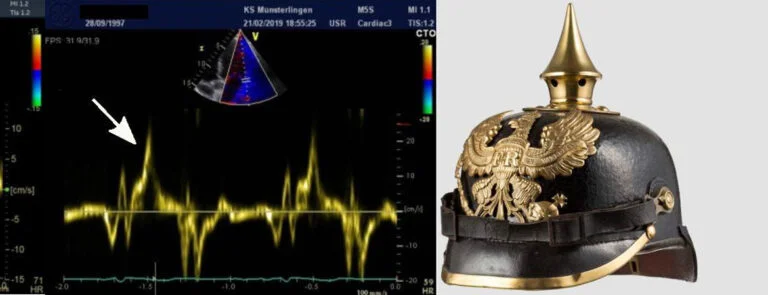
Muthukumar L, et al. The Pickelhaube Sign: Novel Echocardiographic Risk Marker for Malignant Mitral Valve Prolapse Syndrome. JACC Cardiovasc Imaging. 2017 Sep;10(9):1078-1080. doi: 10.1016/j.jcmg.2016.09.016.
Electrocardiography may reveal inferolateral T-wave inversion, QTc prolongation, or frequent premature ventricular complexes. Ambulatory ECG Holter monitoring is recommended to quantify ventricular ectopy and detect non-sustained ventricular tachycardia, while exercise testing may identify exercise-induced ventricular arrhythmias.
Cardiac magnetic resonance imaging has become an essential component of risk stratification in AMVP. CMR enables detection of myocardial fibrosis within the papillary muscles or basal inferolateral left ventricular wall using late gadolinium enhancement and T1 mapping.
Although the genetic basis of AMVP remains incompletely understood, pathogenic variants involving structural proteins, particularly titin (TTN), have been reported in selected patients.
Management
Management of AMVP should be individualized and based on arrhythmic burden, myocardial substrate, and mitral valve morphology rather than on the presence of MAD alone.
Medical therapy is primarily indicated for symptomatic ventricular ectopy, non-sustained ventricular arrhythmias, suspected premature ventricular complex (PVC)-induced cardiomyopathy, or left ventricular systolic dysfunction not fully explained by mitral regurgitation. Beta-blockers and non-dihydropyridine calcium-channel blockers remain first-line agents and provide symptomatic relief, although their effect on reducing PVC burden is generally modest. Class IC antiarrhythmic drugs or amiodarone may be considered in carefully selected patients, balancing antiarrhythmic efficacy against the potential for long-term adverse effects.
Catheter ablation may be considered in patients with symptomatic or high-burden PVCs refractory to medical therapy, PVC-induced cardiomyopathy, or clearly identifiable triggers arising from the papillary muscles or Purkinje system.
Implantable cardioverter-defibrillator (ICD) therapy is recommended for secondary prevention in patients with previous sudden cardiac arrest, ventricular fibrillation, or sustained ventricular tachycardia in the absence of reversible causes. Current evidence does not support routine prophylactic ICD implantation based solely on the presence of MAD or MVP, and decisions regarding primary prevention should therefore be individualized.
Mitral valve surgery is indicated according to current valvular heart disease guidelines in patients with severe degenerative mitral regurgitation accompanied by symptoms or left ventricular dysfunction. In selected patients, surgical correction may also reduce ventricular arrhythmia burden by eliminating the abnormal mechanical forces acting on the papillary muscles and mitral annulus. This potential antiarrhythmic benefit appears greatest in patients with pronounced MAD, excessive annular hypermobility, papillary muscle traction, and limited or absent myocardial fibrosis on CMR.
Lifestyle Recommendations
Patients with AMVP and documented ventricular arrhythmias should avoid high-intensity competitive sports and strenuous resistance exercise, particularly activities such as bodybuilding, as exercise-induced tachycardia and marked increases in blood pressure may augment mechanical stress on the mitral valve apparatus and potentially increase arrhythmic risk. Participation in recreational physical activity should be individualized.
References
1. Piscione M, Pala B, Cribari F, Vignaroli W, Mroue J, Mehta V, Matar F, Perrone MA. The Arrhythmogenic Spectrum of Mitral Valve Disease: Pathophysiology, Risk Stratification, and Surgical Management. J Clin Med. 2026 Jan 21;15(2):865. doi: 10.3390/jcm15020865. PMID: 41598802; PMCID: PMC12841869.
2. Kotyza, V., Segeťová, M., Skalická, B., Peichl, P., Peldová, P., Macek, M., Jr., Kautzner, J., & Krebsová, A. (2026). Arytmogenní prolaps mitrální chlopně, retrospektivní hodnocení možností odhadu rizika život ohrožujících arytmií. Cor et Vasa, 68(3), 308–313. https://doi.org/10.33678/cor.2025.136
3. Chianese S, Ordine L, Pacella D, Canciello G, Di Napoli S, Lombardi R, Esposito G, Losi MA. Arrhythmic risk in mitral valve prolapse with mitral annular disjunction: meta-analysis of longitudinal studies. Sci Rep. 2025 Dec 16;16(1):2428. doi: 10.1038/s41598-025-32285-3. PMID: 41402368; PMCID: PMC12820290.
4. Craig Hacking, Mitral annular disjunction, Radiopaedia.org, Available from: https://radiopaedia.org/articles/mitral-annular-disjunction-1
Authors: Simona Lozinakova, Michal Pazdernik
.jpg)
You Might Also Like
Mitral Annular Disjunction (MAD) And Refractory Ventricular Fibrillation

Eosinophilic Myocarditis Due To Eosinophilic Granulomatosis With Polyangiitis (Churg-Strauss sydrome)