
Introduction
Anterior myocardial infarction has the worst prognosis of all MIs, mostly due to a large size.
The incidence of anterior ST-elevation MI (STEMI) is approximately 33% of all STEMIs.
The anterior wall of the heart is supplied by the left anterior descending artery (LAD). More proximal occlusion is related to larger size of ischemic myocardium.
ST elevations in anterior leads are not the only presentation of anterior ischemia. Other patterns are represented by Wellens syndrome or De Winter T waves.
Etiology
The most common cause of anterior MI is atherosclerotic plaque rupture, followed by thrombus formation and prolonged ischemia in the territory of LAD.
ECG features
- ST elevations in some or all of leads V1 through V6 + I, aVL
- Reciprocal ST depression in the inferior leads (II, III and aVF)
- Septal = V1-2
- Anterior = V2-5
- Anteroseptal = V1-4
- Anterolateral = V3-6, I + aVL
- Extensive anterior / anterolateral = V1-6, I + aVL
ST elevations in leads I, aVL, and V1-V4 and ST depressions in leads II, III, and aVF, suggests the origin of occlusion in the proximal portion of the LAD.
ST elevations in leads V3-V6 and no ST-segment depressions in leads II, III, and aVF, suggests the origin of occlusion in distal part of the LAD.
Management
- coronary angiography/PCI
- UFH
- ASA
- P2Y12 inhibitors
- nitrates
- analgesia
- 02
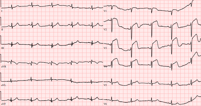
ECG 1 STEMI anterior (ST elevations in V1-V4, ST depressions in inferior leads)
ECG 2 STEMI anterior (ST elevations in V1-V4, ST depressions in inferior leads, RBBB) - proximal LAD occlusion was documented in cath lab.

References
- Bansal K, Gore M, Nalabothu P. Anterior Myocardial Infarction. [Updated 2020 Aug 31]. In: StatPearls [Internet]. Treasure Island (FL): StatPearls Publishing; 2020 Jan-. Available from: https://www.ncbi.nlm.nih.gov/books/NBK562234/
- Tibaut M, Mekis D, Petrovic D. Pathophysiology of Myocardial Infarction and Acute Management Strategies. Cardiovasc Hematol Agents Med Chem. 2017;14(3):150-159. [PubMed]
- Borja Ibanez, Stefan James, Stefan Agewall, Manuel J Antunes, Chiara Bucciarelli-Ducci, Héctor Bueno, Alida L P Caforio, Filippo Crea, John A Goudevenos, Sigrun Halvorsen, Gerhard Hindricks, Adnan Kastrati, Mattie J Lenzen, Eva Prescott, Marco Roffi, Marco Valgimigli, Christoph Varenhorst, Pascal Vranckx, Petr Widimský, ESC Scientific Document Group, 2017 ESC Guidelines for the management of acute myocardial infarction in patients presenting with ST-segment elevation: The Task Force for the management of acute myocardial infarction in patients presenting with ST-segment elevation of the European Society of Cardiology (ESC), European Heart Journal, Volume 39, Issue 2, 07 January 2018, Pages 119–177, https://doi.org/10.1093/eurheartj/ehx393
- https://litfl.com/anterior-myocardial-infarction-ecg-library/

Introduction
Anterior myocardial infarction has the worst prognosis of all MIs, mostly due to a large size.
The incidence of anterior ST-elevation MI (STEMI) is approximately 33% of all STEMIs.
The anterior wall of the heart is supplied by the left anterior descending artery (LAD). More proximal occlusion is related to larger size of ischemic myocardium.
ST elevations in anterior leads are not the only presentation of anterior ischemia. Other patterns are represented by Wellens syndrome or De Winter T waves.
Etiology
The most common cause of anterior MI is atherosclerotic plaque rupture, followed by thrombus formation and prolonged ischemia in the territory of LAD.
ECG features
- ST elevations in some or all of leads V1 through V6 + I, aVL
- Reciprocal ST depression in the inferior leads (II, III and aVF)
- Septal = V1-2
- Anterior = V2-5
- Anteroseptal = V1-4
- Anterolateral = V3-6, I + aVL
- Extensive anterior / anterolateral = V1-6, I + aVL
ST elevations in leads I, aVL, and V1-V4 and ST depressions in leads II, III, and aVF, suggests the origin of occlusion in the proximal portion of the LAD.
ST elevations in leads V3-V6 and no ST-segment depressions in leads II, III, and aVF, suggests the origin of occlusion in distal part of the LAD.
Management
- coronary angiography/PCI
- UFH
- ASA
- P2Y12 inhibitors
- nitrates
- analgesia
- 02
ECG 1 STEMI anterior (ST elevations in V1-V4, ST depressions in inferior leads)
ECG 2 STEMI anterior (ST elevations in V1-V4, ST depressions in inferior leads, RBBB) - proximal LAD occlusion was documented in cath lab.
References
- Bansal K, Gore M, Nalabothu P. Anterior Myocardial Infarction. [Updated 2020 Aug 31]. In: StatPearls [Internet]. Treasure Island (FL): StatPearls Publishing; 2020 Jan-. Available from: https://www.ncbi.nlm.nih.gov/books/NBK562234/
- Tibaut M, Mekis D, Petrovic D. Pathophysiology of Myocardial Infarction and Acute Management Strategies. Cardiovasc Hematol Agents Med Chem. 2017;14(3):150-159. [PubMed]
- Borja Ibanez, Stefan James, Stefan Agewall, Manuel J Antunes, Chiara Bucciarelli-Ducci, Héctor Bueno, Alida L P Caforio, Filippo Crea, John A Goudevenos, Sigrun Halvorsen, Gerhard Hindricks, Adnan Kastrati, Mattie J Lenzen, Eva Prescott, Marco Roffi, Marco Valgimigli, Christoph Varenhorst, Pascal Vranckx, Petr Widimský, ESC Scientific Document Group, 2017 ESC Guidelines for the management of acute myocardial infarction in patients presenting with ST-segment elevation: The Task Force for the management of acute myocardial infarction in patients presenting with ST-segment elevation of the European Society of Cardiology (ESC), European Heart Journal, Volume 39, Issue 2, 07 January 2018, Pages 119–177, https://doi.org/10.1093/eurheartj/ehx393
- https://litfl.com/anterior-myocardial-infarction-ecg-library/


