Endomyocardial fibrosis in a young patient
A 33-year-old female prisoner with a history of asthma bronchiale, sideropenic anemia and methamphetamine abuse was admitted to a hospital for 3 weeks of exertion dyspnea, chest discomfort, dry cough and epigastric pain.
Upon admission CT angio revealed intermediate to high-risk bilateral pulmonary embolism (PE) in lobar arteries, bilateral pleural effusion up to 1 cm and multiple small embolizations to the brain.
Figure 1 CT pulmonary artery angiography confirming bilateral pulmonary embolism


The patient was started on unfractionated heparin (UFH).
Initial echocardiography showed systolic dysfunction of both ventricles (LVEF 30-35%), apical akinesis of fibrotic myocardium and apical obliteration with organized thrombi, right ventricular outflow tract (RVOT) obstruction and circular pericardial separation up to 21 mm in width around right ventricle. Mild mitral and tricuspid regurgitation (1/4), dilated right atrium with two adherent round thrombi. All these findings were pointing towards a diagnosis of endomyocardial fibrosis (EMF).
Video 1 Four chamber view from TEE showing apical obstruction of both ventricles

Video 2 Thrombus in right atrium visible on TEE

Video 3 Severe triangular obstruction of RVOT visible on TEE

Investigation of endomyocardial fibrosis etiology followed with laboratory testing. Eosinophilia was not present at this time and parasitology tests revealed only border-line levels of antibodies against Taenia solium (cysticercosis).
Elevation of marker Ca-125 could mean a paraneoplastic etiology, but whole body CT scan showed only mild hepatomegaly and ascites with no other sign of malignant processes.
The only other promising etiologic factor could be vasculitis with positive anti ds-DNA and ANCA anti-MPO antibodies. Patient was referred to a rheumatology department for further investigation.
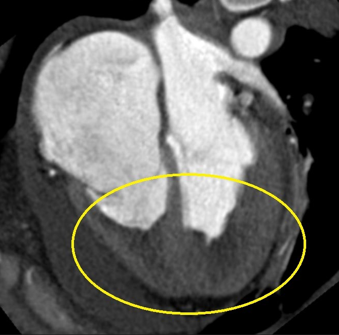
Other imaging techniques were performed to further define the tissue characteristic of the intracardiac masses and the underlying cardiac tissue. Cardiac MR could not be unfortunately performed for patient’s claustrophobia and non-cooperation. CT angio of the heart confirmed changes similar to endomyocardial fibrosis with intracardiac thrombi in both ventricles and right atrium, bilateral PE, pericardial and pulmonary effusion.
Figure 2 Cardiac CT confirming apical obstruction of both ventricles

On the 10th day the patient developed heparin induced thrombocytopenia (HIT) and unfractionated heparin (UFH) was changed to fondaparinux (Arixtra) in combination with acetylsalicylic acid (ASA).
Intracardiac echocardiography with endomyocardial biopsy (EMB) of the intracardiac mass from the right ventricle was performed on the 22nd day since hospitalization. Histology confirmed the presence of organized thrombi. Pericardiocentesis of 250 ml slightly green fluid was performed at the same time and sent for laboratory testing, cultivation was negative. Therapy with Ibuprofen and Colchicine was started with good results.
Video 4 ICE navigated biopsy of mass in right ventricle

Social investigation of this young prisoner showed a problematic situation (debts, execution and severe non-compliance), therefore the patient was not indicated for surgical decortication of the ventricles. She was started on medical therapy for chronic heart failure and anticoagulation and transferred for further investigation of possible ANCA+ vasculitis to the Rheumatology centre in a different institution.
Discussion
Endomyocardial fibrosis (EMF) is an idiopathic disorder that is characterized by fibrosis of the apical endocardium of the right ventricle (RV) and/or left ventricle (LV). This leads to the development of restrictive cardiomyopathy and diastolic dysfunction with increased filling pressures. It is also typically associated with mitral or tricuspid regurgitation, because the chordae tendineae are often involved. This leads to atrial enlargement, thrombus formation over the initial lesions and congestive heart failure.
EMF is mostly endemic to the tropical and subtropical regions of the world, but sporadic forms may be found in Europe. The European form is associated with a mean age of 30–50 years and a male-to-female ratio of 1:2.
What is the cause of EMF?
EMF has an idiopathic pathophysiology, but is often associated with an excessive blood eosinophilia, which may result from a parasite infection (helminths, protozoa – malaria, toxoplasmosis), some autoimmune diseases (vasculitis, rheumatoid arthritis, post-transplant rejection) or eosinophilic leukaemia. The significance of eosinophilia is still controversial, but it is believed that the eosinophils are mechanically destroyed in the ventricles, which releases fibroblast-stimulating factors that cause the typical lesions in the inflow tract and apex.
Nevertheless, despite the similarities between Loeffler’s endocarditis and EMF, serum and myocardial eosinophilia have not been consistently demonstrated in EMF.
Other causes include infectious origin, environmental exposure, immunologic or genetic.
The development of EMF may be divided into 3 stages:
- acute carditis phase - eosinophilic infiltration of the myocardium with subendocardial necrosis, characterized by a febrile illness and in severe cases cardiogenic shock. Pericardial effusion may be present.
- sub-acute phase – a decrease in the amount of inflammatory activity, associated with thrombus formation over the initial lesions.
- chronic fibrotic phase - the endocardium is replaced by collagenous fibrosis. It is characterized by chronic HF with restrictive physiology and symptoms as ascites, peripheral oedema and cardiomegaly accompanied by typical echocardiographic findings, including apical fibrosis and restrictive ventricular filling pattern. Usually, there is no eosinophilia present in later stages of disease.
Echocardiography is the main imaging method for EMF diagnosis.
- Apical fibrosis of the right ventricle (RV), left ventricle (LV), or both ventricles, apical thrombi are often present
- Tethering the atrioventricular (AV) valve papillary muscles, leading to mitral and/or tricuspid regurgitation
- Restrictive ventricular filling pattern
- A pericardial effusion is frequently present and may be large.
- Atrial enlargement, often with adherent thrombi.
There is a triad of key features of endomyocardial fibrosis:
- Apical obliterations of one or both ventricles (echocardiogram or angiocardiogram).
- Mild cardiomegaly with severe pulmonary congestion.
- Mitral and/or tricuspid regurgitation; diastolic heart failure.
Cardiac magnetic resonance (CMR) - imaging with contrast demonstrates myocardial fibrosis
What can usually be found on ECG?
- atrial fibrillation – 1/3 of patients with EMF
- low QRS voltage due to myocardial fibrosis
- AV blocks
- right or left bundle branch blocks
- ventricular arrhythmias – fibrotic lesions in ventricular myocardium
Management of endomyocardial fibrosis
Medical care currently remains very challenging as 1/3 to 1/2 of patients with advanced disease die within 2 years from diagnosis. Clinical course may be stable over years, but when restriction becomes clinically apparent, a downhill course begins with a decrease in quality of life. For patients with severe symptoms, consider surgical therapy because the prognosis for these patients with continued medical therapy alone is dismal.
Medical treatment
- If the patient presents in early stages with acute myocarditis, corticosteroids may prevent disease progression and necrotizing myocarditis.
- Heart failure treatment for diastolic dysfunction – diuretics and rate control for atrial fibrillation or flutter (digitalis, beta-blockers) are currently the mainstays of therapy. ACE inhibitors and ARBs may prove useful with their antifibrotic properties, but there are currently no data to support this yet.
Surgery
- Endocardial decortication seems to be beneficial for many patients with advanced endomyocardial fibrosis (EMF) who are NYHA III or IV. The operative mortality rate is high (15-20%), but successful surgery offers the possibility of long-term survival when the remaining myocardium is not severely fibrotic and dysfunctional.
- Typically, one or both ventricles are decorticated, with mitral or tricuspid valve replacement because of involvement of chordae tendineae and papillary muscles.
- After decortication most patients show an increase in cardiac volumes.
Prognosis
The prognosis for EMF is poor as the incidence of sudden cardiac death due to arrhythmia, thromboembolic disease, and end-stage heart failure is very high
What can you include in differential diagnosis?
- Anthracycline toxicity
- Carcinoid heart disease
- Fabry disease
- Fatty infiltration
- Glycogen storage disease
- Gaucher disease
- Hurler disease
- Idiopathic cardiomyopathy
- Metastatic cancers
- Rheumatic heart disease
- Radiation
References
- Ali A Sovari, M. (2020, December 29). Endomyocardial fibrosis. Retrieved May 09, 2021, from https://emedicine.medscape.com/article/154931-overview
- CAMM, A. J., LÜSCHER, T. F., & SERRUYS, P. W. (2009). The ESC textbook of cardiovascular medicine. Oxford, Oxford University Press
- Shaaheen Laher, Yee Weng Wong, David Platts, Anil Prabhu, Bruce Thomson, Christian Hamilton-Craig, David Godbolt, Elizabeth Cheesman, Alexander Dashwood, A Rare Case of Severe Nontropical Isolated Right Ventricular Endomyocardial Fibrosis, JACC: Case Reports, Volume 2, Issue 13, 2020, Pages 2078-2084, ISSN 2666-0849, https://doi.org/10.1016/j.jaccas.2020.09.042. (https://www.sciencedirect.com/science/article/pii/S2666084920311979)
- Bhatti K, Bandlamudi M, Lopez-Mattei J. Endomyocardial Fibrosis. [Updated 2020 Aug 12]. In: StatPearls [Internet]. Treasure Island (FL): StatPearls Publishing; 2021 Jan-. Available from: https://www.ncbi.nlm.nih.gov/books/NBK513293/
- Rebecca Cogswell, MD, Nelson B Schiller, MD, FACC, FRCP, FASE, Harry Acquatella, MD, FACC, FAHA (2020). Endomyocardial fibrosis. In Todd F Dardas, MD, MS (Ed.), UpToDate. Retrieved May 10, 2021 from: https://www-uptodate-com.ezproxy.is.cuni.cz/contents/endomyocardial-fibrosis?search=endomyocardial%20fibrosis&source=search_result&selectedTitle=1~111&usage_type=default&display_rank=1
Authors: Lucie Mayerová, Michal Pazderník

You Might Also Like
.gif)
Hemolytic anemia after mitral valve repair

Quantification of cardiac shunt (atrial septal defect)